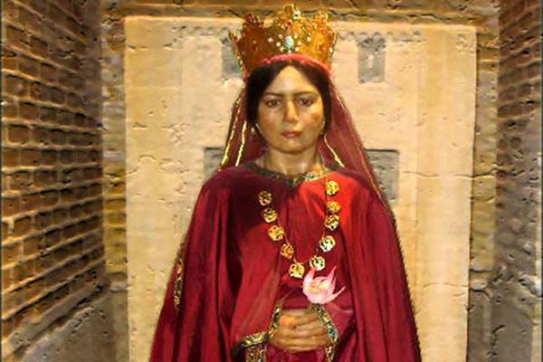
The first recorded case of an inflammatory breast cancer: Persian empress Atossa (550 BC to 475 BC), daughter of Cyrus the Great, wife of two Achamenian kings, Cambyses and Darius.
“Cancer may have started the fight, but I will finish it.”
Introduction
Since cancer-prevention is not possible, the saying, “prevention is the cure” is amended to “early detection is the cure.”
Only about 10% of cancer deaths are because of primary tumour. Most of the deaths are because of metastasis – spreading of the cancer to other parts of the body. Once metastasis happens, it is difficult to treat the cancer. Early detection of cancer is therefore of utmost importance.
Early Detection
Several ways of early detection:
- Self-examination of Breasts
More than 80% cancers are detected by women doing self-examination of breasts. The examination should be done every month, 5-7 days after menorrhoea. Look for the following:
- Lumps in breast (less than 20% are cancer) or in lymph nodes in armpits.
- Thickening of breasts
- One breast becoming larger than other
- A nipple changing position or shape or becoming inverted
- Discharge from nipple
- Constant pain in part of breast or armpit
- Swelling beneath the armpit or around the collarbone
‘Breast cancer self-check’ images and videos are available on internet. In case of palpated anomaly, consult your gynecologist.
The limitations of self-examination are:
- Only 20% women do self-examination of breasts.
- The tumour/changes are large by the time they are felt and this delay in detection can adversely affect the treatment outcome.
- Imaging Techniques
Early detection of cancer is required and is possible by using Imaging Techniques. Six Imaging Techniques are available:
- X-rays examination. Small neoplasmatic tissue formations can be seen.
- Sonography (Ultrasound)
Sonography is done in addition to Mammography to rule out possible cysts and to estimate the size of the tumour. However, tumours smaller than 5 mm cannot be detected.
- MRI
MRI is used to detect if the breast has been affected by more than one tumour.
- Computer Assisted Detection (CAD)
CAD is used to point out possibly diseased regions. It is used mainly as a second opinion to the report of the doctor.
- CT-scan
CT-scan is most often used to see if breast cancer has spread to other organs.
- PET
A PET scan is used to detect the cancer cells in the body. It is often combined with a CT scan (known as a PET/CT scan).
Limitations of Imaging
- Imaging techniques magnify the tumour much as the magnifying glass magnifies the letters in a book. If the font size is very small, a letter cannot be identified even with the magnifying glass. In a similar way, the imaging techniques cannot identify tumours that are very small.
- The QUALITY of cancer is more important than the QUANTITY. A small tumour can be more dangerous than a large tumour. Imaging can tell the quantity of the tumour, that is, its size, but cannot tell the quality of the tumour.
- Most of the time, Imaging cannot even tell whether a tumour is cancerous or not.
Confirming Cancer
The only absolute way to confirm cancer is by biopsy: a small tissue from the tumour is taken and microscopically examined to check for cancer.
Types of Biopsy
- Punching Biopsy. Done in a locally sedated state.
- Needle Biopsy. Done with a syringe and a special needle. As painful as venepuncture.
- Advanced Breast Biopsy Instrumentation (ABBI). Done with X-ray to ensure localisation of target. Only a few doctors are experienced in this technique.
Microscopic examination of biopsy is sufficient; but in a few rare cases specialized lab tests are required.
Treatment
Even small localised tumours have the potential of metastasis and therefore need to be treated. The treatment is surgery, medications (hormonal therapy and chemotherapy), radiation and immunotherapy.
Surgery offers the single largest benefit. Used along with chemotherapy and radiation, the local relapse rate is reduced, and the overall survival rate may increase.
Surgery
- Mastectomy: remove whole breast.
- Quadrantectomy: remove quarter breast.
- Lumpectomy: remove small part of breast.
- Endoscopy-assisted breast-conserving surgery (EBCS), which has the advantage of a less noticeable scar, was developed more than ten years ago.
- Breast Reconstruction Surgery or breast prostheses: to simulate breast.
Neo-adjuvant, that is prior to surgery, and Adjuvant that is after and in addition to surgery, medication is used as part of treatment. For example, Neo-adjuvant use of aspirin may reduce the mortality from Breast Cancer.
Adjuvant Therapies
Radiation (negative effect on normal cells) to kill cancer cells in tumour bed and regional lymph nodes that may have escaped surgery. It reduces the risk by 50 – 66 % (i.e., 1/2 to 2/3 reduction of risk). It is confined to region being treated. But only solid tumour can be treated.
Therapies using drugs/agents etc.
- Chemotherapy (negative effect on normal cells). Uses drugs, usually two or more drugs in combination, to destroy cancer cells.
- Targeted Therapy that became available in 1990s that uses drugs that inhibit enzymes.
- Monoclonal Antibody Therapy in which the agent is an antibody
- Immunotherapy that uses patient’s immune systems to fight cancer using drugs.
- Hormone Blocking Therapy. Uses Estrogen Receptors (ER +) Tamoxifen and Progesterone Receptors (PR +) Anastrozole that block the receptors.
Experimental Cancer Treatment
- Gene Therapy
- Ultrasound Energy.
Complementary and Alternative Cancer Treatments
Alternative cancer treatments do not cure cancer. But they may lessen signs and symptoms – such as anxiety, fatigue, nausea and vomiting, pain, difficulty sleeping, and stress – caused by cancer and cancer treatments. Hypnosis, massage, meditation, relaxation techniques, yoga, acupuncture, aromatherapy, music therapy, and Tai chi – alone or in combination may be beneficial.
But many alternative cancer treatments are unproved, and some may even be dangerous.
Type of Treatment Given
Patients with good prognosis are offered less invasive treatment – e.g. lumpectomy + radiation + hormone.
Patients with poor prognosis are offered more aggressive treatment – extensive mastectomy + radiation + chemotherapy + adjuvant medication.
Treatment Success Rate
If the cancer is detected early, that is at Stage 1, prognosis is excellent and usually chemotherapy is not required.
If detected in Stage 2 & 3 prognosis is progressively poorer with a greater risk of recurrence. Surgery, chemotherapy, and radiation are required.
If detected in Stage 4, that is metastatic cancer (spread to distant sites), prognosis is poor. Surgery, radiation, chemotherapy, and targeted therapies are used. But the 10-year survival rate is 5% without treatment and 10 % with optimal treatment.
In India, more than 60% of the BC’s are diagnosed at stage III or IV. Hence the low survival rate.
Psychological and Emotional Aspects
Cancer patients need psychological and emotional support. Besides the family, such support can be given by support groups who are trained and experienced in giving such support. ‘Cancer Sahyog’ is one such support group in India.
Conclusion
Cancer is a 3200-year-old disease. It is endogenous, a part of life-process. So, it can neither be eradicated, nor prevented, nor cured. As yet.
Late detection of cancer is fatal. The causes for late detection are many but lack of awareness is the principal cause. Other main causes are patient being shy, social stigma and doctors’ ignorance because of which the treatment is delayed. An awareness program will address all these issues.
Will some radical discovery in the future make cancer prevention and cure possible? We do not know. But we can always hope.
Because as Richard Causer, Director, NCI, USA, says about the future of cancer cure, “There are far more good historians than there are prophets.”